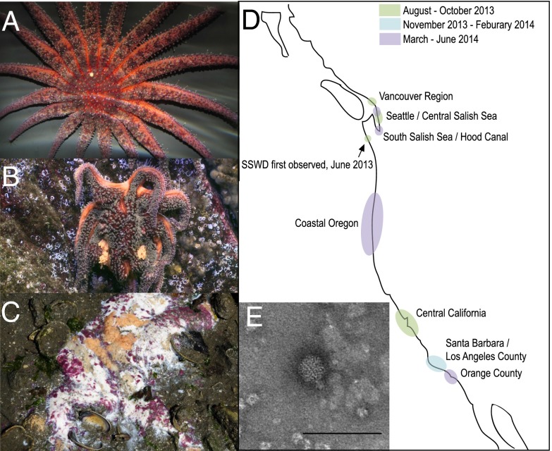
Fig. 1.
Photographs of SSWD-affected stars (A) asymptomatic P. helianthoides, (B) symptomatic P. helianthoides, and (C) symptomatic P. ochraceus. Disease symptoms are consistent with loss of turgor, loss of rays, formation of lesions, and animal decomposition. (D) Map showing occurrence of SSWD based on first reported observation. (E) Transmission electron micrograph of negatively stained (uranyl acetate) viruses extracted from an affected wild E. troschelii from Vancouver . The sample contained 20–25-nm diameter nonenveloped icosohedral viral particles on a background of cellular debris (primarily ribosomal subunits) and degraded viral particles of similar morphology. (Scale bar: 100 nm.)

