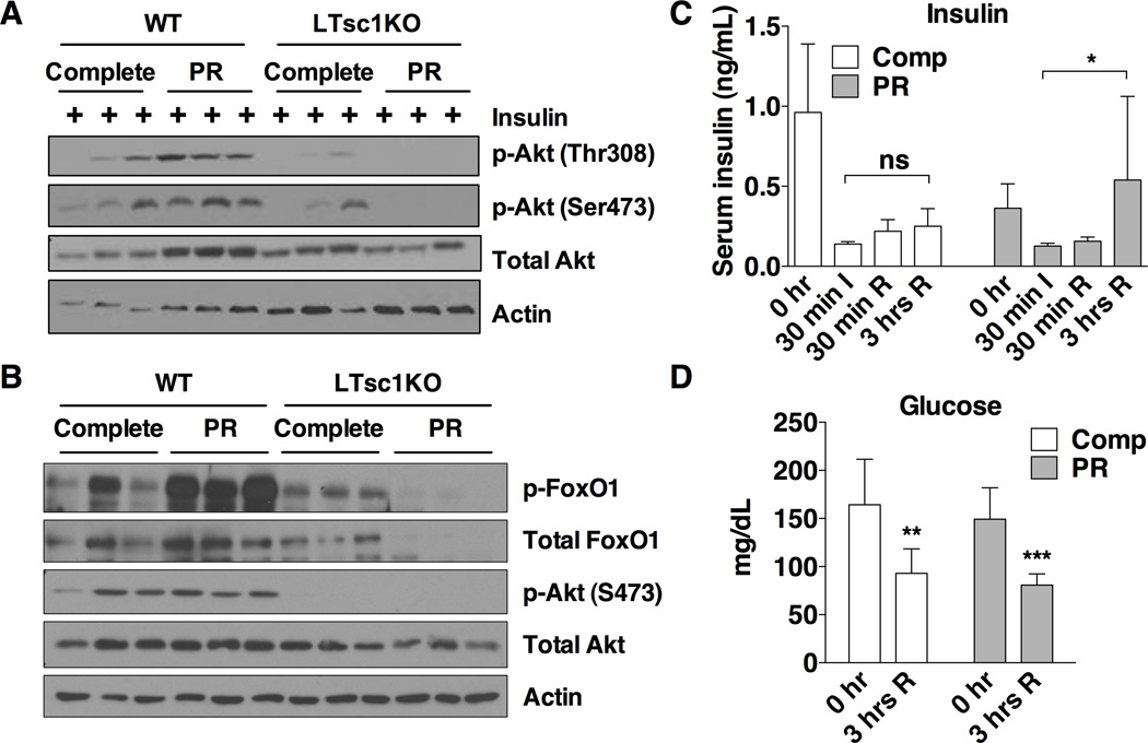
Figure 5. The TSC complex is required for improved hepatic insulin sensitivity upon PR.
(A) Insulin sensitivity as determined by immunoblotting for markers of Akt pathway activation in liver extracts from mice fasted for 6 hrs and then stimulated with 0.5 U/kg insulin by portal vein injection 3 min before harvest.
(B) Akt activation status as determined by immunoblotting of liver extracts 3 hrs after reperfusion from mice preconditioned for 1 wk on the indicated diet prior to induction of hepatic IRI.
(C) Serum insulin levels from tail blood of WT mice preconditioned for 1 wk on the indicated diet taken prior to ischemia (0 hr), at the end of the ischemic period (30 min I, n=3–4), 30 min after reperfusion period (30 min R, n=3) or 3 hrs after reperfusion (3 hrs R, n=11–12). Asterisk indicates the significance of the indicated comparison according to a Kruskal-Wallis test followed by Dunn’s multiple comparisons test; *p < 0.05; ns: not significant.
(D) Blood glucose levels of mice on the indicated diets for 1 wk before hepatic IRI, measured before (0hr) and 3 hrs after reperfusion; n = 8–10 mice/group. Asterisks indicate the significance of the difference between 0 and 3 hr values by student’s t test within diet group; **p < 0.001, ***p < 0.0001.
Data in all panels are shown as means ± SD. See also Figure S5.

