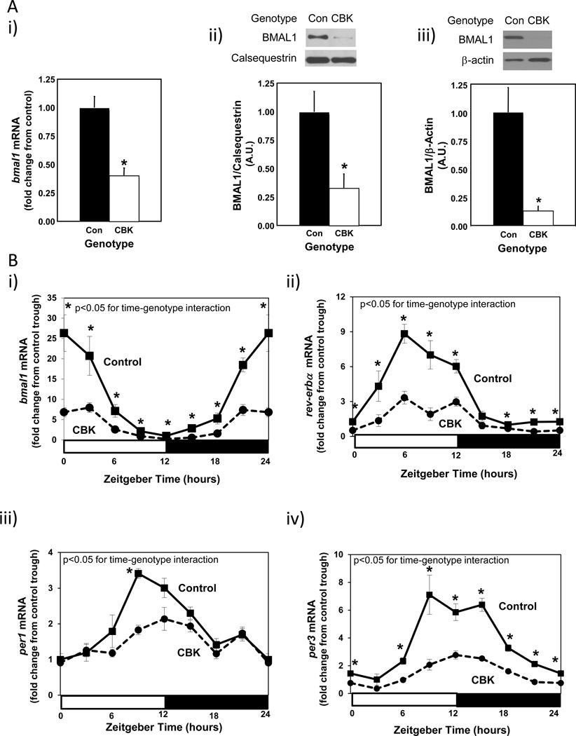
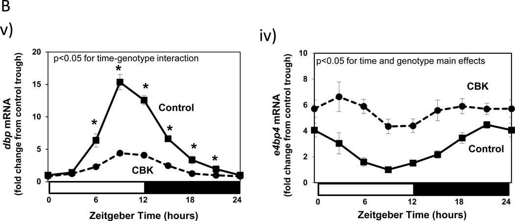
Figure 1.
Decreased bmal1 mRNA levels in intact CBK hearts (Ai), decreased BMAL1 protein levels in intact CBK hearts (Aii), and in cardiomyocytes isolated from CBK hearts (Aiii), as well as attenuated time-of-day-dependent gene expression oscillations in circadian clock components (bmal1, rev-erbα, per1, and per3) and clock output genes (dbp and e4bp4) in CBK hearts (B). Mice were housed in a 12-hr light:12-hr dark cycle (lights on at ZT0). Hearts were isolated from 12 week old male CBK mice and age-matched littermate controls. For data presented in A, hearts were isolated at ZT6. Note that ZT0 and ZT24 are identical data. Values are expressed as mean ± SEM (n=4–12; sample size range varies dependent on the parameter investigated). *, p<0.05 for CBK versus littermate control at a distinct ZT (post-hoc pair-wise comparison).


