
Figure 2. Phosphorylation of p130Cas, c-Src and JNK downstream of integrin αv following mechanical stimulation.
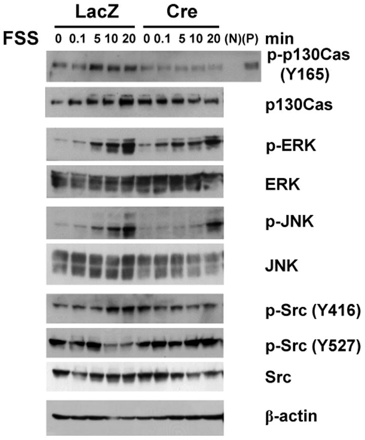
Integrin av-deficient osteoblasts show impaired phosphorylation of p130Cas and JNK in response to FSS. Calvaria-derived primary osteoblasts from αv flox/flox mice were infected with adeno-LacZ (-) or adeno-Cre (+) vectors, and subjected to FSS. Total cellular protein was isolated from control (LacZ) and integrin αv-deficient (Cre) osteoblasts at 0, 0.1, 5, 10, and 20 min following FSS, and immunoblotted with the indicated antibodies. N and P indicate negative and positive controls for phosphorylated p130Cas (p-p130Cas), which represent unattached cells in suspension and phenylarsine oxide-treated attached cells, respectively.