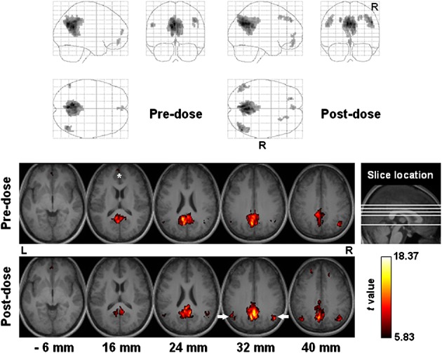
Figure 5.

Functional connectivity of the default mode network among all subjects (n = 17). Top panel: projection viewed from right (sagittal), behind (coronal, right side on the right) and above (axial, right side at bottom). Bottom panel: sectional axial view in neurological convention. Slice location is marked in units of millimeters. The asterisk and arrows indicate the medial prefrontal cortex and lateral parietal cortex, respectively.
