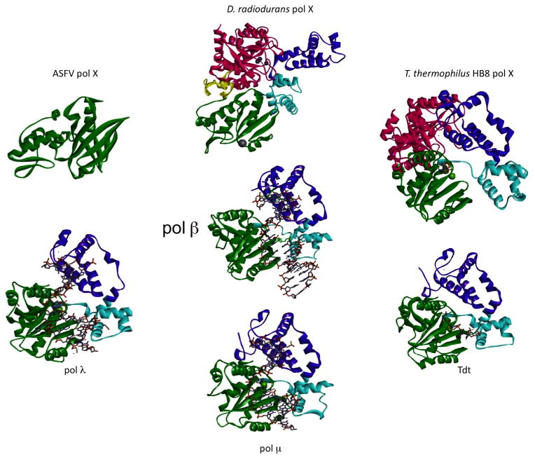
Fig. 5.
Representative crystal structures of X-family DNA polymerases. The domains/subdomains are colored according to the scheme used in Fig. 4 and are arranged to show similar orientations. The PDB IDs are: human pol β, 2FMS [60]; ASFV pol X, 1JAJ [16]; D. radiodurans pol X, 2W9M [13]; T. thermophilus HB8 pol X, 3AU2 [45]; truncated mouse Tdt, 1KDH [61]; truncated mouse μ, 2IHM [62]; truncated human pol λ, 2BCR [63]. When present, DNA is shown in a stick representation.

