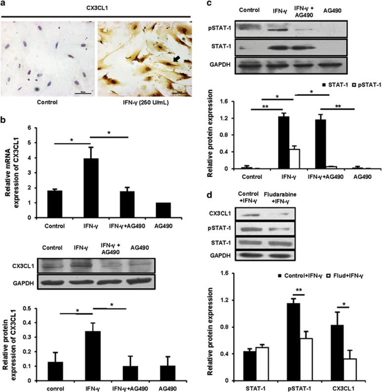
Figure 4.
IFN-γ upregulated CX3CL1 via a JAK2-STAT1 pathway in uterine stromal cells. (a) The isolated uterine stromal cells were treated with or without IFN-γ at a dose of 250 U/ml for 12 h, and CX3CL1 protein expression was analysed by immunocytochemical staining. The arrow indicates that CX3CL1 protein expression is markedly induced in response to IFN-γ. Scale bar: 100 μm. (b) Uterine stromal cells were pretreated with AG490 at 10 μM for 2 h before IFN-γ treatment, and then CX3CL1 expression was analysed by quantitative PCR (top panel) and western blotting (bottom panel). Data show the mean±S.E.M. of three independent experiments, respectively. *P<0.05 by one-way analysis of variance (ANOVA). (c) The treatment was the same as described in (b). The STAT1 and pSTAT1 were analysed by western blotting and normalized to GAPDH and STAT1, respectively. Data show the mean±S.E.M. of three independent experiments. *P<0.05 and **P<0.01 by one-way ANOVA. (d) Uterine stromal cells were pretreated with fludarabine at 100 μM for 2 h before IFN-γ treatment, and then CX3CL1, pSTAT1 and STAT1 were analysed by western blotting. CX3CL1, STAT1 and pSTAT1 were normalized to GAPDH, GAPDH and STAT1, respectively. Data show the mean±S.E.M. of three independent experiments. *P<0.05 and **P<0.01 by independent samples T-test

