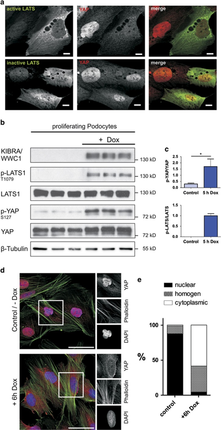
Figure 5.
LATS-dependent reactivation of Hippo pathway in podocytes results in a nuclear export of YAP. (a) Transient expression of a permanent active LATS2 mutant (T1041E, green) and a permanent inactive LATS2 mutant (T1041A, green) each tagged with a FLAG epitope in podocytes led to an increase of cytoplasmic YAP (red) localization in cells expressing the permanent active mutant but not the permanent inactive mutant. Scale bars represent 10 μm. (b) Indirect activation of LATS1 kinase due to inducible KIBRA/WWC1 overexpression (doxycycline treatment, 125 ng/ml for 5 h) was used to verify the link between LATS activation and nuclear export of YAP. Cell lysates of doxycycline-treated and -untreated cells were utilized to detect endogenous LATS1, p-1079-LATS1, YAP, p-S127-YAP, KIBRA/WWC1 and β-tubulin, which served as loading control. Overexpression of KIBRA/WWC1 in podocytes results in a robust increase in LATS and YAP phosphorylation. (c) The ratio between phosphorylated and total amount of the protein is calculated and indicated (mean and S.D., t-test,*P<0.05). (d) Immunofluorescence staining of YAP (red) in podocytes after induction of the KIBRA/WWC1 overexpression by doxycycline for 6 h: overexpression of KIBRA/WWC1 leads to a cytoplasmic localization of YAP; Phalloidin (green) marks the actin cytoskeleton, DAPI (blue) was used to visualize nuclei. Scale bars represent 50 μm. (e) Quantitative evaluation of 100 cells shown in c proves the general translocation of YAP. For each cell, the localization of YAP was estimated as nuclear, cytoplasmic or homogeneous (comparable levels of nuclear and cytoplasmic staining)

