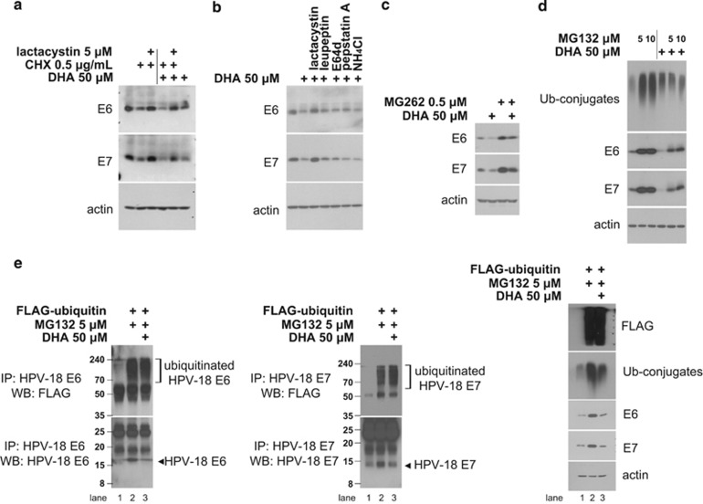
Figure 2.
DHA induces the UPS-dependent degradation of E6/E7 viral oncoproteins. (a) HeLa cultures were pretreated, or not, with lactacystin (5 μM) for 1 h before the addition of CHX (0.5 μg/ml), DHA (50 μM) or a combination of the two. Cells were collected 6 h later and subjected to western blot analysis. (b) HeLa cells were left untreated or treated with 50 μM DHA for 6 h with 1 h of pretreatment of 5 μM lactacystin, 10 μM leupeptin, 2.5 μg/ml E64d, 1 μM pepstatin A and 10 mM NH4Cl, respectively. Whole-cell lysates were extracted and blotted with antibodies against E6/E7. (c) HeLa cells were preincubated with or without 0.5 μM MG262 for 1 h before the addition of 50 μM DHA. After 6 h, whole-cell lysates were blotted with anti-E6/E7 antibodies. (d) HeLa cells were pretreated, or not, with 5 μM or 10 μM MG132 for 1 h and then treated with 50 μM DHA for 6 h. The expression levels of indicated proteins were then examined by western blotting (WB). (e) DHA increases E6/E7 ubiquitination. HeLa cells transiently expressing a control vector or FLAG-ubiquitin plasmid were pretreated, or not, with 5 μM MG132 for 1 h and then treated with 50 μM DHA for 6 h. Whole-cell lysates were subjected to immunoprecipitation (IP) with anti-E6 (left) or -E7 (middle) antibodies, as indicated, followed by WB with anti-FLAG (top) or anti-E6/E7 (bottom) antibodies. Right, whole-cell lysates were blotted with anti-FLAG, -ubiquitin (Ub-conjugates) and -E6/E7 antibodies to examine expression levels

