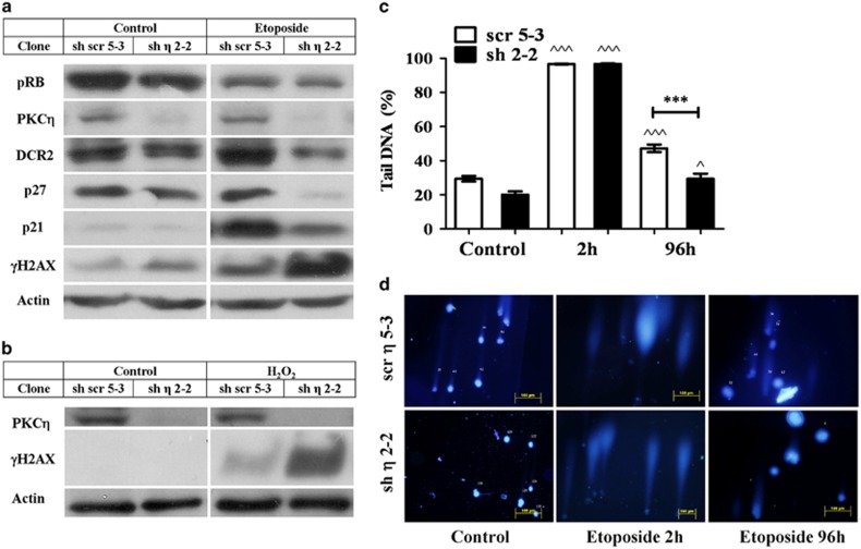
Figure 2.
PKCη-knockdown reduces senescence markers. MCF-7 clones expressing sh-PKCη (sh2-2) or scrambled control cells (scr 5-3) were treated with (a) 400 μM etoposide or (b) 150 μM H2O2 for 2 h. Fresh medium was added for 96 h followed by cell lysis. The senescence markers p21Cip1, p27Kip1, DcR2 and pRB, along with the DDR marker γ−H2AX were detected using Western blot analysis and specific antibodies. β-Actin was used as a marker for equal protein loading. The results shown are representative of three independent experiments. (c) Average percentage of DNA in comet tail. Cells were treated by etoposide as described above, detached by trypsin at the indicated time points, counted and subjected to single-cell electrophoresis as described in Materials and Methods. Results are the average of the volumes of comet tails of at least 250 cells per time point (A minimum of 50 different fields were photographed in three separate experiments). Error bars represent the S.D.; two-tailed, unpaired sample t-test statistical analysis is shown: ^ indicates statistical significance compared with untreated cells of the same clone, * indicates statistical significance compared with other clones. ^P≤0.05 and ^^^/***P≤0.0001. (d) Shown are representative pictures of comet assay analyses of sh-PKCη and scrambled control clones at indicated time points

