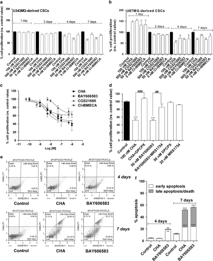
Figure 2.
Effects of AR agonists on CSC proliferation. U343MG-derived CSCs (a) or U87MG-derived CSCs (b) were treated for the indicated number of days with the A1AR agonist (CHA), the A2AAR agonist (CGS21680), the A2BAR agonist (BAY606583) or the A3AR agonist Cl-IBMECA at selected concentrations (corresponding to tenfold the values of the affinity constants). (c) CSCs derived from U343MG cells were incubated for 7 days with the AR agonists at increasing concentrations. (d) CSCs derived from U343MG cells were incubated for 7 days with 100 nM CHA, in the absence or in the presence of the A1AR antagonist DPCPX (50 nM), or with 50 nM BAY606583, in the absence or in the presence of the A2BAR antagonist MRS1754 (20 nM). At the end of the treatments, cell proliferation was evaluated using the MTS assay. The data were expressed as a percentage with respect to that of untreated cells (control), which was set to 100%, and they are the mean values±S.E.M. of three independent experiments, each performed in duplicate. (e and f) CSCs were treated for 4 or 7 days with NSC medium containing DMSO (control), 100 nM CHA or 50 nM BAY606583. At the end of treatments, the cells were collected and the degree of phosphatidylserine externalisation was evaluated using the Annexin V protocol, as described in the Materials and method section. (f) The data were expressed as the percentage of apoptotic cells (early-stage apoptotic cells shown in white, late-stage apoptotic/necrotic cells shown in grey) relative to the total number of cells. The data shown are the mean values±S.E.M. of three different experiments. The significance of the differences was determined using a one-way ANOVA with the Bonferroni post-test. *P<0.05, **P<0.01, ***P<0.001 versus control. ##P<0.01, ###P<0.001 versus agonist alone

