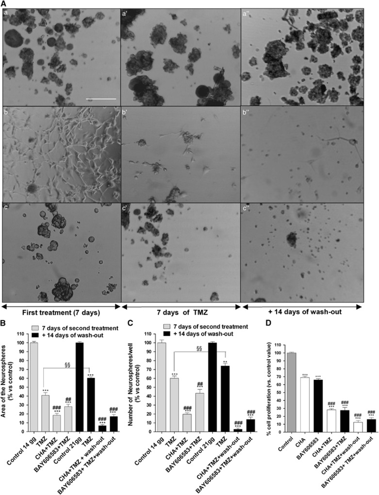
Figure 8.
Effect of the sequential treatment of CSCs with an A1AR or A2BAR agonist and TMZ on their morphology. (A) CSCs were treated for 7 days with complete NSC medium containing DMSO (control) (a), 100 nM CHA (b) or 50 nM BAY606583 (c); after 7 days, the cells were treated for another 7 days with complete NSC medium containing DMSO (a') or 100 μM TMZ (b', c'). At the end of treatment periods, the drug-containing media were replaced with fresh drug-free NSC medium, and the cells were cultured for another 14 days (a”, b”, c”). Representative micrographs taken after 7 days of treatment and after 7 or 14 days of drug wash-out are shown A. The area of the culture plates occupied by the spheres (B) and the number of spheres (C) were determined after 7 days of treatment and after 7 and 14 days of drug wash-out. The counts are the mean values±S.E.M. of three independent experiments. The significance of the differences was determined using a one-way ANOVA with the Bonferroni post-test: **P<0.01, ***P<0.001 versus control; ##P<0.01, ###P<0.001 versus TMZ alone. (D) CSCs were treated as in A and their proliferation was evaluated using the MTS assay. The data were expressed as the percentages relative to that of the untreated cells (control), which was set at 100%, and they are the mean values±S.E.M. of three independent experiments, each performed in duplicate. The significance of the differences was determined using a one-way ANOVA with the Bonferroni post-test: ***P<0.001 versus control; ###P<0.001 versus single-agent-treated cells. §§P<0.01 versus cells treated for seven days with TMZ

