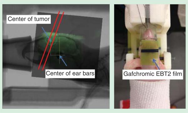
Figure 5.
Left: A 2D registered image of MR image and X-ray radiograph, with three microbeams planned to irradiate across the tumor area; Right: a picture of U87 glioma-bearing mouse fully stabilized on the homemade holder with ear bar and teeth fixation. Gafchromic EBT2 film was used at the microbeam entrance and exit plane for dosimetry confirmation purposes.

