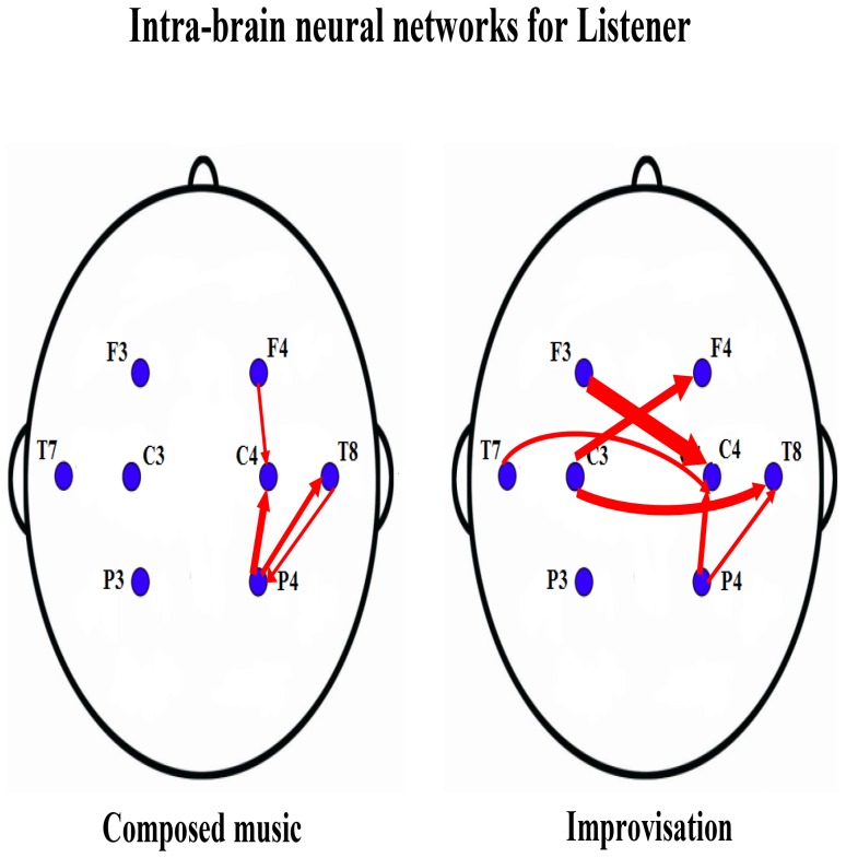
Figure 2. Listener's intra-brain neural networks for the first experiment.
The two panels show the listener's intra-brain neural networks separately for composed music (left) and improvisation (right). The large brain regions are labeled by the 8 electrodes: F3, F4, C3, C4, T7, T8, P3, P4. The red links indicate the direction of neural information flow between large brain regions, where the thickness of the links represents the magnitudes of the causalities.

