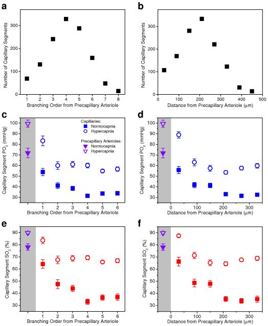
Figure 4. Number of capillary segments and capillary oxygen content as a function of branching order and distance from the precapillary arterioles.
(a, b) Histograms of the capillary segment number. (c-f) PO2 and SO2 inside the capillary segments. Solid and empty symbols in c-f represent measurements during normocapnia and hypercapnia, respectively. Average PO2 and SO2 values in precapillary arterioles (purple triangle symbols) were plotted inside the shaded regions on the left hand side of the graphs. At each branching order and distance from precapillary arteriole, mean PO2 and SO2 during hypercapnia experiments are statistically larger than mean PO2 and SO2 during normocapnia (Student t-test, P<0.006). Data are expressed as mean ± s.e.m. and were estimated averaging over all vessels over all animals. n = 6 mice were used for (a) and (b) and n = 3 mice were used for each group in (c-f).

