
Figure 8. Simplified representation of the cortical microvascular path, intravascular oxygenation, and supplied tissue territories.
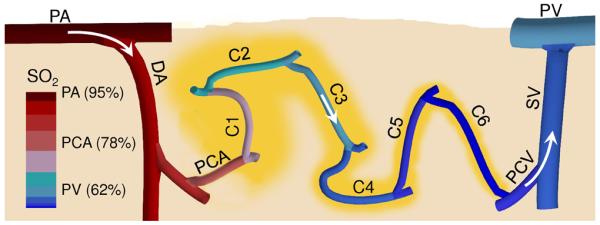
A vertical cross section through the cortical tissue revealing one microvascular path close to the cortical surface including pial arteriole (PA), diving arteriole (DA), precapillary arteriole (PCA), capillary path (C1-C6) with the low branching order capillaries (C1-C3) and high branching order capillaries (C4-C6), postcapillary venule (PCV), surfacing venule (SV), and pial venule (PV). Arrows represent the direction of the blood flow. Vascular segment colors represent approximate average segment SO2. Tissue territories supplied by capillary segments were outlined in yellow.