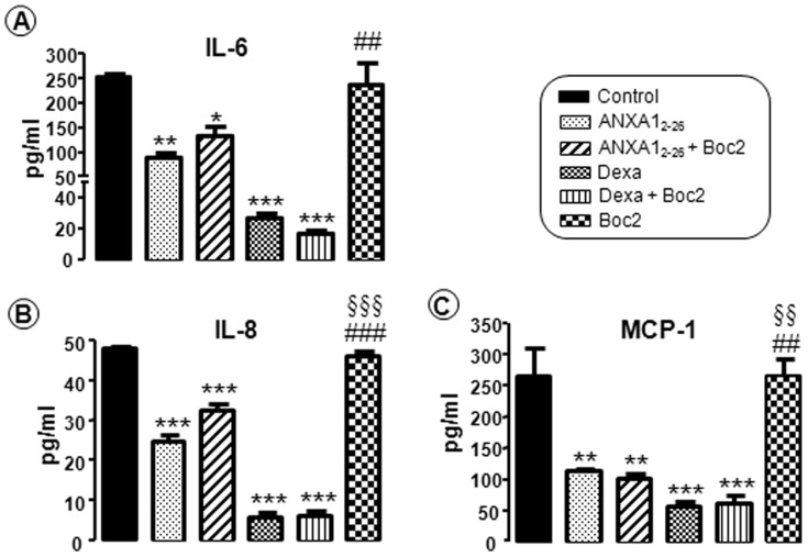
Figure 5. Effect of the peptide ANXA12–26 on proinflammatory cytokine expression.
Low expression of IL-6 (A), IL-8 (B) and MCP-1 (C) after treatment with ANXA12–26 and dexamethasone. Hep-2 cells were seeded in MEM-Earle medium at a density of 2×10⁶ cells in 75-cm2 culture flasks, and then were incubated with serum-free medium 24 hours prior to the addition of ANXA12–26 (1 µM), ANXA12–26 (1 µM)+Boc2 (10 µM), Dexa (0.01 µM), Dexa (0.01 µM)+Boc2 (10 µM) or Boc2 (10 µM) alone. All of the experiments were performed in triplicate to confirm the results. Data are expressed as the mean ± SEM of the analyte concentration (pg/mL), determined using MAGPIX xPONENT software. * P<0.05, ** P<0.01 and *** P<0.001 vs. control, ## P<0.01 and ### P<0.001 vs. ANXA12–26, §§ P<0.01 and §§§ P<0.001 vs. ANXA12–26+Boc2.

