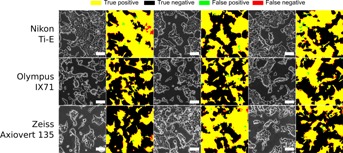
Figure 3.

Comparison of segmentation performance for images of a single Oct4-GiP mESC culture acquired using different phase contrast microscopes, cameras and imaging protocols. The microscopes used were a Nikon Ti-E microscope (Fi-1 color camera), a Olympus IX71 (Hamamatsu ORCA-ER C4742-80-12AG monochrome camera) and a Zeiss Axiovert 135 (Hamamatsu ORCA-R2 C10600-10B-H monochrome camera). Each row is a different microscope. Three fields of view per microscope were considered. The raw phase contrast microscopy image is shown with the segmentation result overlaid in white. Next to it is the comparison with the manually annotated ground truth image. All processing parameters were kept constant. Scale bars are 100 µm.
