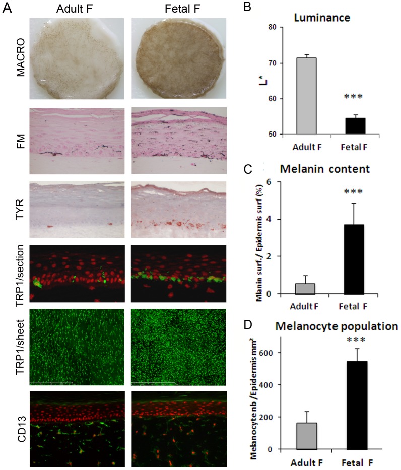
Figure 2. Comparison of the effect of fibroblasts from fetal versus adult origin on reconstructed skin pigmentation.
PRS samples were reconstructed with either fetal fibroblasts (GM10) or adult (21 yr-old donor) fibroblasts within the dermal equivalent. Identical keratinocyte and melanocyte strains were used for the epidermal reconstruction. A drastic increase in pigmentation of the PRS and activation of melanocytes in the presence of fetal fibroblasts as compared to adult fibroblasts, were noted A) macroscopically (Macro), on histological sections stained with Fontana-Masson (FM) and by tyrosinase staining (Tyr) of tissue sections. The hyper-pigmentation observed in fetal versus adult fibroblast condition was quantified by B) a decrease in Luminance value and C) an increase in melanin content. TRP-1 labeling of tissue sections showed that melanocytes were correctly located at the basal layer in both conditions (A). TRP-1 staining of epidermal sheets revealed an increase in melanocyte numbers in the presence of fetal fibroblasts (A) which was confirmed by image analysis (D). CD13 staining revealed no change in fibroblast morphology or density (A). Values are expressed as the mean +/- SD calculated for 4 different samples in 2 independent experiments and analyzed using the two-tailed unpaired Student's t-test, *** p<0.001. Magnifications (A): FM, TYR, and TRP-1/section = x400, CD13/section = x200, TRP1/sheet = x50.

