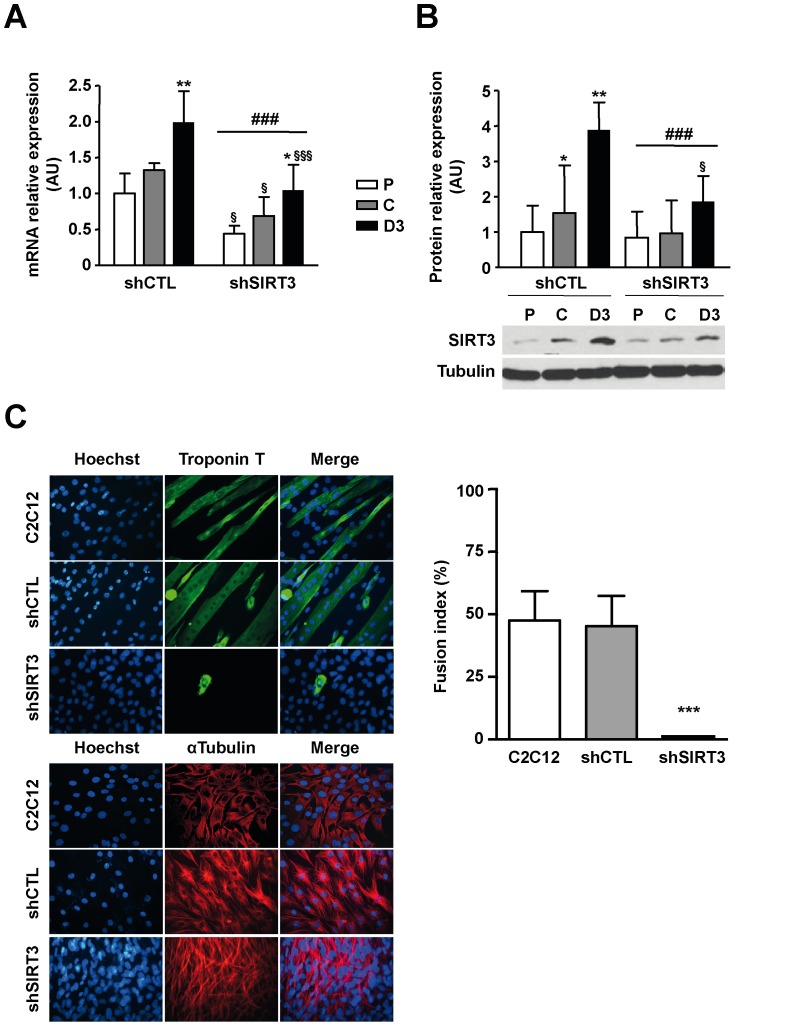
Figure 2. Depletion of SIRT3 impairs terminal myoblast differentiation.
Endogenous SIRT3 expression in C2C12-LucshRNA (shCTL) and C2C12-SIRT3shRNA (shSIRT3) cells during proliferation (P), at cell confluence (C) and after 3 days of differentiation (D3). A) SIRT3 mRNA expression was monitored by real-time RT-PCR and normalized relatively to the reference genes ARP and TBP. Results are expressed as the mean ± SD of three independent experiments. B) Western blot analysis of SIRT3 protein expression. Quantification was performed with Image J software and normalized relatively to Tubulin protein level. Results are expressed as the mean ± SD of three separate experiments. ANOVA main effect: ### P<0.001 vs. shCTL cells. Post-hoc significance: *P<0.05, **P<0.01 and ***P<0.001 vs. proliferating myoblasts for each cell type. §P<0.05, §§P<0.01 and §§§ P<0.001 vs. shCTL cells at the same statstatee. C) Immunostaining of C2C12, C2C12-LucshRNA (shCTL) and C2C12-SIRT3shRNA (shSIRT3) cells with an anti-Troponin T (differentiated state) or anti α-tubulin (undifferentiated state) antibody and fusion index 3 days after the induction of differentiation. Nuclei were stained with Hoechst. Microphotographs of a typical experiment are shown (x400). Fusion index values are expressed as the mean ± SD of 10 images/dish using image J software. *** P<0.001 vs. shCTL cells.

