
Fig. 1.
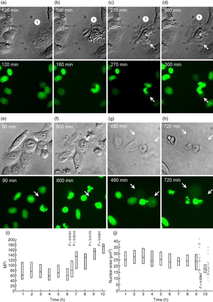
Redistribution of histone 2B-green fluorescent protein (H2BGFP) during ultraviolet (UV)-B-induced apoptosis in HeLa cells. Adherent HeLa cells expressing fluorescent H2BGFP histone were cultured to a density of 3 × 10⁴ cells/cm2. After UV-B irradiation with 900 mJ/cm2 the distribution of H2BGFP was monitored by video-microscopy. (a–d) The characteristic nuclear changes that had been described for adherent cells undergoing apoptosis. After 270 min the cell marked with ‘1’ had undergone waves of formation of apoptotic cell-derived membranous vesicles (ACMV). None of these contained detectable amounts of H2BGFP; 30 min later two of the vesicles were filled with substantial amounts of H2BGFP (arrows). Note that (1) not all ACMV contain degraded chromatin (H2BGFP); (2) ACMV are formed initially without nuclear material, and are loaded in a second step (arrows). (e,f) Some large ACMVL remain unloaded even 10 h after their formation. (g,h) In very late phases of apoptosis some ACMVL which had been loaded with H2BGFP disintegrate and release their load into the culture supernatant, most probably during secondary necrosis. (i) mean fluorescence intensity (MFI) of the nuclei during the 10 h after irradiation. Note a significant increase of the MFI starting 6 h after irradiation (Student's t-test of cellular MFI at t = 0 versus t = × hours after irradiation). (j) nuclear area during the 10 h after irradiation. Note a significant shrinkage of the nuclei 9 h after irradiation (Student's t-test of cellular nuclear area at t = 0 versus t = × hours after irradiation).