Fig. 1.
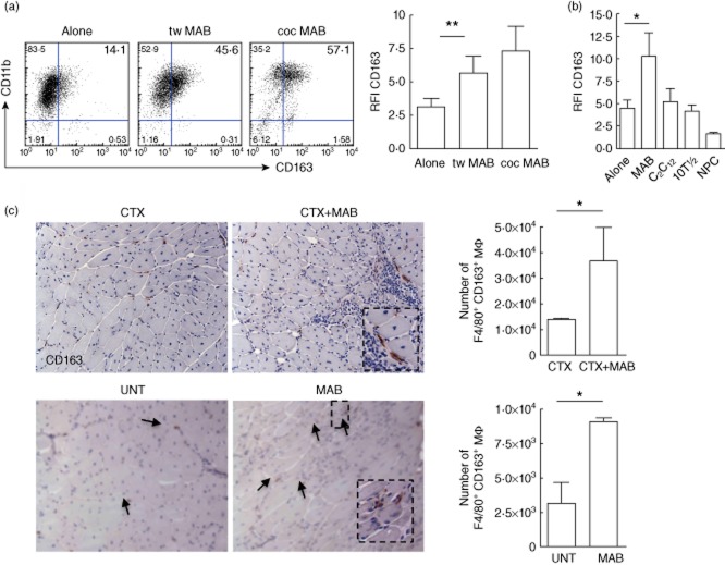
Mesoangioblasts regulate macrophage CD163 expression. Macrophages cultured for 48 h alone (alone), or challenged with mesoangioblasts (MAB) either in culture conditions allowing physical cell-to-cell contact (co-culture, coc) or separated by a semi-permeable membrane (Transwell, Tw) were analysed by flow cytometry for CD163 expression. (a) Representative dot-plots indicating frequency of CD11b+CD163+ cells and CD163 relative fluorescence intensity (RFI). Error bars indicate the mean ± standard error of the mean (s.e.m.) of 11 independent samples. **P < 0·01. (b) CD163-associated fluorescence (RFI) was assessed in macrophages cultured alone or challenged with MAB, C2C12 immortalized myoblasts, 10T1/2 embryonic fibroblasts or neural precursor cells (NPC). Error bars indicate the mean ± s.e.m. of three independent experiments. *P < 0·05. (c) CD163 expression was assessed by immunohistochemistry: in the skeletal muscle of C57Bl6 mice in which mesoangioblasts had been transplanted (CTX+MAB) or not (CTX) 24 h after injury (top panels); in the skeletal muscle of C57Bl6 αSG−/− dystrophic mice (bottom panel) transplanted (MAB) or not (untreated, UNT) with mesoangioblasts. (d) Mononuclear cells were retrieved after enzymatic digestion of the skeletal muscle. F4/80+CD163+ macrophages were identified within CD45+CD11b+ leucocytes by flow cytometry. Error bars indicate the mean ± s.e.m. of three independent experiments with three mice per group. *P < 0·05.

