
Fig. 2.
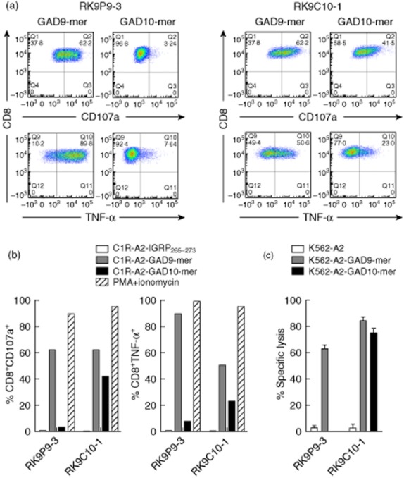
Differential recognition of peptide-pulsed human leucocyte antigen (HLA)-A*0201+ cell lines by CD8 T cell clones. Representative intracellular staining for tumour necrosis factor (TNF)-α and surface staining of CD107a (degranulation marker) of clone cells after 18 h co-culture with peptide-pulsed target cell lines. (a) RK9P9-3 shows a high percentage of events positive for TNF-α production and degranulation following co-culture with glutamic acid decarboxylase 9 (GAD9)-mer-pulsed target cells, but minimal positivity using GAD10-mer-pulsed target cells. In contrast, RK9C10-1 displays a cross-reactive profile to both GAD9-mer- and GAD10-mer peptide-pulsed cell lines. (b) These responses contrasted against a negative control peptide stimulation (IGRP265–273) and the positive control of phorbol myristate acetate/ionomycin stimulation. (c) Specific lysis of GAD9-mer-pulsed K562-A2 target cells by both RK9P9-3 and RK9C10-1 and killing of GAD10-mer-pulsed target cells by RK9C10-1 only; 10 000 live CD3+CD8+ events were recorded for flow cytometry analyses; cytotoxicity and functional assays are representative data performed at an effector : target ratio of 25:1 for 4 h. Data shown are representative single experiments.