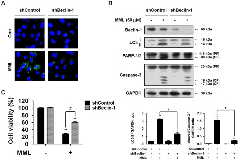
Figure 5. Knockdown of Beclin-1 gene suppresses MML-induced apoptotic cell death.
(A) Control and Beclin-1 knockdown cells were treated with MML (60 µM) for 24 h and then LC3 immunofluorescence staining was carried out for detecting autophagosomes. Nuclei were stained with DAPI. Images were captured by confocal microscope (bar; 10 µm). (B) Knockdown cells (shControl, shBeclin-1) were treated with MML (60 µM) for 24 h. Western blot analysis was performed with antibodies against Beclin-1, LC3, PARP-1/2 and caspase-3, respectively. GAPDH was used as loading control. Bar graphs indicate the ratio of LC3-II/GAPDH and cleaved caspase-3/GAPDH, respectively. (C) Control and Beclin-1 knockdown cells were treated with MML (60 µM) for 24 h and then cell viability was measured by MTT assay. Bar graph represents the percentage of cell viability. All data were expressed as mean ± SEM of three independent experiments. *p<0.05 and **p<0.01 compared with control; # p<0.05 compared with MML-treated group.

