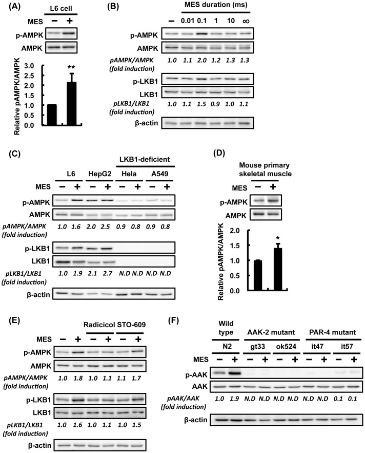
Figure 5. MES induces AMPK activation via LKB1-dependent pathway.
(A) Differentiated L6 cells were treated with MES (0.1 ms, 1 V/cm, 55 pps) for 10 min. Cell lysates were extracted 2 hr after MES treatment. Phospho-AMPK (Thr-172) and AMPK were detected by Western Blotting analysis. Relative amount of p-AMPK was normalized to total AMPK. Data are presented as mean ± SE (n = 3). **P<0.01 versus control, assessed by unpaired Student t-test. (B) Differentiated L6 cells were treated with MES (1 V/cm, 55 pps) at various pulse widths (0.01, 0.1, 1 and 10 ms) or in the absence of pulse (∞ ms) for 10 min. Cell lysates were extracted 2 hr after MES treatment, and analyzed using the indicated antibodies. (C) Differentiated L6, HepG2, Hela and A549 cells and (D) mouse primary skeletal muscle cells were treated with MES (0.1 ms, 1 V/cm, 55 pps) for 10 min. Cell lysates were extracted 2 hr after MES treatment, and analyzed using the indicated antibodies. (E) Differentiated L6 cells were pre-treated with radicicol (10 µM, 1 hr) or STO-609 (10 µM, 1 hr) and then co-treated with MES for 10 min. Cell lysates were extracted 2 hr after MES treatment. Proteins were subjected to Western blotting analysis using the indicated antibodies. (F) Worms were treated once a day with MES for 20 min at larval stage. Worm lysates were extracted 2 hr after MES treatment, and analyzed by Western blotting using the indicated antibodies. β-actin served as internal control. For (A–F), blots shown are representative of 2-3 independent experiments. The number under each blot is the intensity of the blot relative to that of untreated control. N.D., not-detected due to too low intensity.

