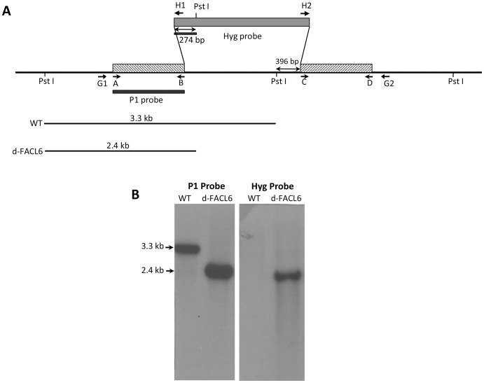
Figure 5. Generation of FACL6-deletion mutant of M. tuberculosis.
(A), Schematic depiction shows the genomic locations of the primers and probes used in the construction and confirmation of facl6 deletion mutants. The sequences of the primers are given in Table 1. (B), Genomic DNA from WT Mtb and d-facl6 mutant was digested with PstI and hybridized with the 5′-flank of the d-facl6 construct as probe and the hyg probe. Wild-type genomic DNA digested with PstI and probed with the 5′ flank of the disruption construct yielded a hybridization fragment of 3.3 kb (lane WT). In contrast, PstI digested DNA from the mutant strain showed a smaller band of 2.4 kb due to the presence of a PstI site in the 5′ region of the hyg cassette. Hybridization with the hyg probe showed the expected band in the mutants and no hybridization with the WT DNA.

