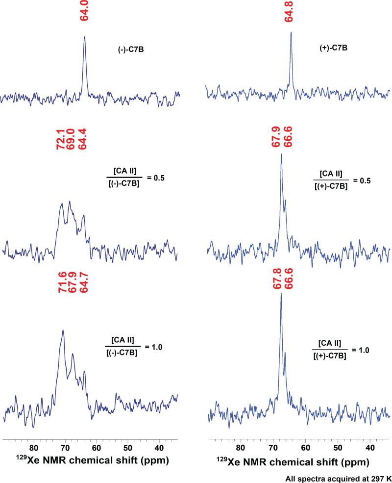
Figure 2.
HP 129Xe NMR spectra of (−)-C7B (left column) or (+)-C7B (right column) before and after binding to wild-type carbonic anhydrase II. The first row shows spectra of biosensors (138 μM, (−)-C7B; 147 μM (+)-C7B) dissolved in 90% Tris-SO4 buffer (50 mM, pH 8.0) and 10% glycerol. The second and third rows show spectra for biosensor-CA II binding. Solutions of 1.3 mM CA II in 50 mM Tris-SO4 buffer were titrated with 0.5 and 1.0 equivalents of biosensor, respectively.

