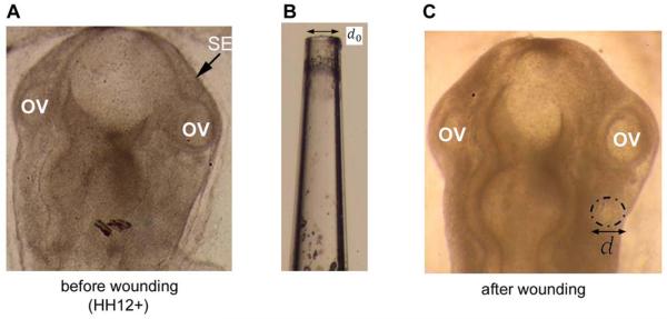
Figure 3.
Tension estimate in surface ectoderm (SE). (A) Dorsal view of HH12+ chicken embryo before wounding. OV=optic vesicle. (B) Glass needle used to make hole in surface ectoderm. Internal diameter is d0 = 90 μm. (C) Same embryo immediately after wounding SE. Dash-dot line outlines the wound, which is relatively circular with a diameter d larger than the pipette tip (d = 97 ± 3.0 μm, n = 8), indicating nearly isotropic tension.

