Figure 8.
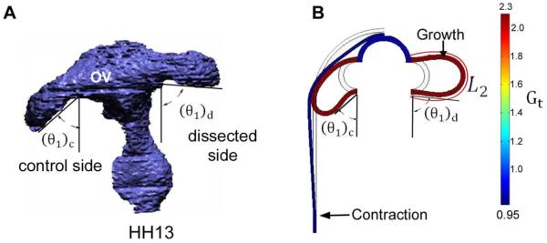
Computational model for effects of surface ectoderm (SE) on optic vesicle (OV) shape (ventral view). (A) 3-D reconstruction after dissection of SE from one OV (stage HH13). After removing SE, the OV angle θ1 increased relative to the control side. The caudal bending angle in this plane, defined as φ1 = 90 − θ1, decreased from 33 ± 8° on the control side to 5 ± 4° on the dissected side (n = 5). (B) Frontal-plane model at HH13 for control case (left side) and without SE (right side). Uniform tangential growth is specified in OV with isotropic contraction in SE. The bending angle is 42° on the control side and 8° on dissected side. L2 = circumferential length of OV.

