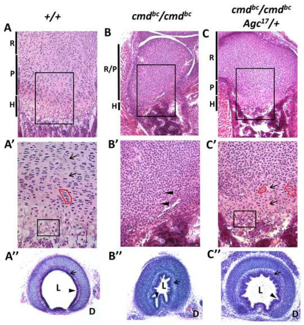
Figure 3. Histological examination of mutant and rescue growth plate cytoarchitecture.
Representative sections of 18.5 dpc femoral growth plates stained with hematoxylin and eosin demonstrating chondrocyte morphology within each growth plate zone (A–C) for wild-type, mutant, and rescue embryos. (A’–C’) Higher magnification of the proliferative and hypertrophic regions outlined in black boxes in top row. Red outlines highlight the columnar formation of proliferative chondrocytes, and the black box denotes chondrocytes with hypertrophic morphology, both of which are absent in aggrecan-null growth plates. Note the hypercellularity and lack of extracellular matrix in mutant embryos (arrow heads) compared to wild type and rescue (arrows). (A’’–C’’) Sections of cartilaginous rings from the upper third of the trachea, showing differences in airway patency with and without aggrecan. R= resting, P= proliferative, H= hypertrophic, L= lumen, D= dorsal aspect.

