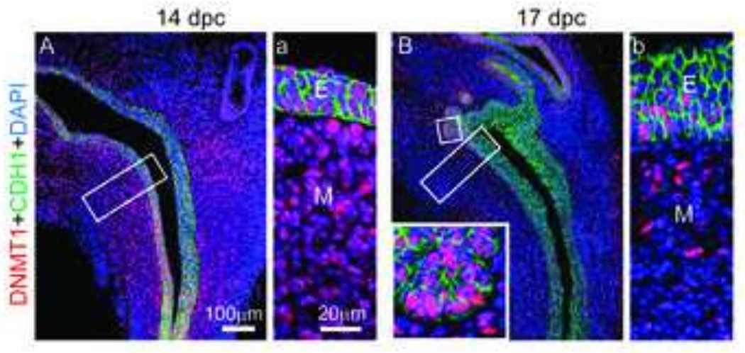
Figure 1. DNMT1 protein abundance diminishes in the mesenchyme during prostate development.
(A,a) 14 dpc and (B,b) 17 dpc lower urinary tract sagittal sections were stained by immunohistochemistry to visualize DNMT1 (red) protein and all epithelium E-cadherin (CDH1, green). Cell nuclei were stained with DAPI. Insets represent magnified images. Abbreviations: E, epithelium; M, mesenchyme.

