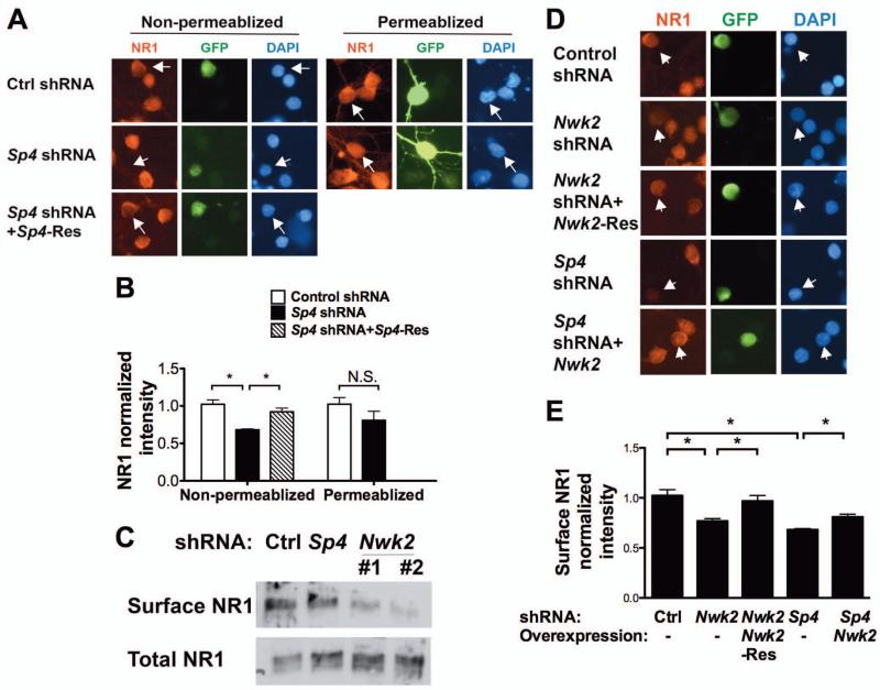
Figure 4. Sp4 regulates NMDAR1 surface levels via Nwk2.
(A,B) CGNs were transfected with plasmids expressing Sp4 shRNA and GFP and, where indicated, RNAi-resistant Sp4 (Sp4-Res). 5 days post-transfection, non-permeabilized or permeablized neurons were immunostained with antibody against NR1. Transfected cells were identified by GFP autofluorescence (non-permeabilized) or staining with anti-GFP (permeabilized). (A) Representative images of neurons are shown. Arrowheads indicate the transfected neurons. (B) Quantification of fluorescence intensity of NR1 immunostaining at the cell body normalized to untransfected neurons in the same field. (C) CGNs infected with lentivirus expressing Sp4 or Nwk2 shRNA and GFP were selected (Supplemental Figure 4) and cell surface proteins labelled with Biotin and purified. Total cell lysates (10%) and surface proteins were analysed by immunoblot with anti-NR1. (D, E) CGNs were transfected with the indicated plasmids and GFP. NR1 immunostaining in non-permeabilized cells was analysed as above. (D) Representative images of neurons are shown. Arrowheads indicate the transfected neurons. (E) Quantification of fluorescence intensity of NR1 immunostaining at the cell body normalized to untransfected neurons in the same field. (B and E) Values represent mean ± SEM of an experiment repeated 3 times and analysed by ANOVA followed by a post-hoc Tukey test; *P<0.05, n.s.= not significant.

