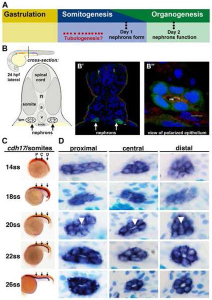
Figure 1. Renal progenitors exhibit a progression of rearrangements during nephrogenesis, and undergo tubulogenesis with lumen formation between the 20-22 ss time points.
(A) Although the timing of tubulogenesis during pronephros development has been unresolved, we hypothesized that it transpires sometime during somitogenesis, as prior studies have demonstrated that nephron patterning is completed by embryonic day 1, and that pronephros function initiates at approximately day 2. (B) The zebrafish embryonic kidney is comprised of two bilateral nephrons that have a lumen at 24 hpf. (B’) Cross section of a zebrafish embryo in which the nephron tubule epithelial cells were labeled by IF to detect GFP (green) in the transgenic strain Tg(cdh17:eGFP), along with acetylated α-tubulin (light blue), Prkcι/ζ (red), and nuclei marked with DAPI (dark blue). (B”) Digital zoom of a single nephron tubule, white bar indicates 5 um. (A-B”) Schematics and images adapted with author rights (Gerlach and Wingert, 2013). (C) Renal progenitors were labeled in the developing pronephros between the 14 and 26 ss by performing WISH on wild-type embryos for the renal marker cdh17 (purple) and a somite marker (red) to confirm embryo stage (the somite marker myod1 was used embryos <18 ss and smyhc1 for embryos >18 ss). Black arrows indicate approximate region where proximal (P), central (C), or distal (D) cross-sections were analyzed. (D) Serial sections of WISH labeled embryos were collected and analyzed at proximal, central, and distal regions of the renal progenitor field. At 14 ss, renal progenitors were found in clusters of approximately ~8 cells. Between 18-22 ss, renal progenitors were found in circumferential clusters of ~6 cells. At the 20 ss, a small lumen was discernible (white arrowheads), and a clear lumen was subsequently visible in all regions of the pronephros at 22 ss that enlarged by 26 ss.

