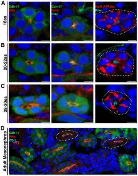
Figure 2. Changes in protein localization precede lumen formation in the pronephros, and similar polarity features characterize embryonic and adult zebrafish nephron tubules.
Localization of proteins during nephrogenesis in wild-type transgenic Tg(cdh17:eGFP) zebrafish embryos and in the uninjured adult kidney. Tubules not labeled with GFP (due to incompatibility of particular antibody-fixative combinations) are outlined with white dots. (A) At the 18 ss: Prkcι/ζ and actin localized to the apical membrane domain prior to the appearance of a lumen, while Na+/K+ ATPase surrounded the cell membrane and was not excluded from the apical surface. (B) When a lumen formed at the 20-22 ss: Prkcι/ζ encompassed the apical membrane domain abutting the lumen, actin was found in a punctate pattern at the apical surface presumably localizing to junctional complexes, and Na+/K+ ATPase still surrounded a majority of the cell membrane but was not present in the region where Prkcι/ζ was localized. (C) At the 28-30 ss: Prkcι/ζ demarcated the apical membrane, actin was more band-like at the apical surface yet still exhibited punctate expression at cell junctions, and Na+/K+ ATPase showed a distinct basolateral membrane localization. (D) In the adult kidney, or mesonephros, Prkcι/ζ was located at the apical surface of tubule cells, and the tight junction marker ZO-1 showed strong labeling at junctional complexes. (A-C) Scale bars, 5 μm. (D) Scale bar, 20 μm.

