
Figure 1.
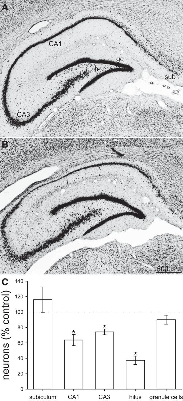
Nissl-stained hippocampi from a control (A) and epileptic pilocarpine-treated (B) rat. sub, Subiculum; h, hilus; gc, granule cells. C, Number of neurons in the subiculum, CA1 cell layer, CA3 cell layer, hilus, and granule cell layer of the dorsal hippocampus. Values represent mean ± SEM of epileptic rats (n = 13) relative to average values of controls (n = 4). *p < 0.05, two-tailed t test.