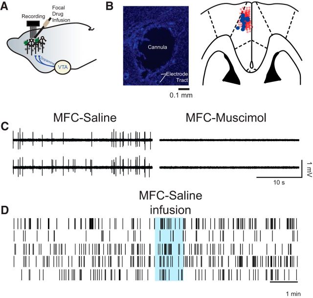
Figure 2.
Medial frontal cortex (MFC) infusions. A, We stereotaxically implanted cannula at a high angle to target recording electrodes in MFC. B, Photomicrograph of a cannula tract and immediately neighboring electrodes (left; section stained with DAPI). Cannula locations were reconstructed from histological sections (right); the cannula is marked by blue circles, and electrode locations are marked by light red circles. C, Wide band (unfiltered signal) from two electrodes in B showing single units in saline sessions and no spiking activity in sessions with muscimol infused into the MFC. Across 135 electrodes in nine animals, we could not identify any single units in sessions with muscimol infused into MFC. D, Saline infusion did not change neural activity or our ability to isolate neurons during an acute recording session.

