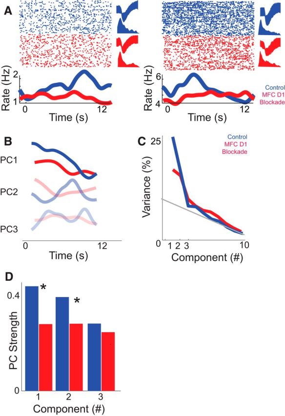
Figure 6.

Medial frontal cortex (MFC) D1DR blockade decreases ramping activity. A, Examples of MFC ramping neurons recorded in control (blue) sessions. In red, the same putative neuron from the control session is shown in MFC D1DR blockade sessions (red). These neurons were identified based on similar waveforms (top right) and interspike intervals (bottom right) in each condition. Note that these are two exemplars. The raster plot at the top shows neuronal activity as each dot represents an action potential (top); each row is a trial. The bottom line plot displays the average firing rates over time. Statistical comparisons assumed that independent populations were recorded in control and D1DR blockade sessions. B, C, Principal component analysis in control sessions (blue) revealed that ramping activity was the most prominent pattern of neural activity among MFC neurons (PC1; B) and explained 27% of variance (C). In MFC D1DR sessions (red), a consistent ramping component was not observed, and PC1 explained less variance. D, To directly compare ramping activity, we projected PCs from control sessions onto MFC D1DR sessions. PC1 explained significantly less variance in MFC D1DR blockade sessions, whereas PC3 was unchanged. Asterisks represent significance at p < 0.05 via a t test. Together, these data suggest that focal D1DR blockade attenuates ramping activity of neurons in MFC.
