
Figure 7.
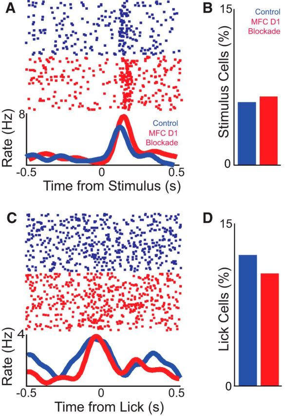
Medial frontal cortex (MFC) D1DR blockade does not change task-related modulations. A, A putative neuron in control and D1DR blockade sessions showed identical stimulus-onset modulations. As in Figure 6, these are exemplars, and statistical comparisons assumed that independent populations were recorded in control and D1DR blockade sessions. The top portion displays the activity of a single neuron, and each dot represents an action potential; each row is a trial. The bottom line plot displays the average firing rates over time. B, A similar fraction of neurons were stimulus onset modulated in control and MFC D1DR blockade sessions, as computed via a paired t test of firing rate 100 ms before/after stimulus onset; note the different time scale compared with Figure 6A. C, A single putative neuron in control and D1DR blockade sessions showed similar lick-related modulation, as computed via a paired t test of firing rate 100 ms before/after lick. D, The percentage of lick-modulated cells was also similar in control and MFC D1DR blockade sessions. Significance was determined at a level of p < 0.05.