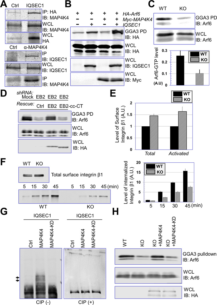
Figure 6. MAP4K4 promotes IQSEC1 and Arf6 activity.
(A) Interaction between IQSEC1 and MAP4K4 was confirmed by coimmunoprecipitation. Cells were transfected with plasmids encoding Myc-tagged MAP4K4 with or without HA-tagged IQSEC1. Cell lysate was immunoprecipitated with α- HA and blotted with different antibodies as indicated (top panel). To verify interaction of endogenous proteins, lysate of WT keratinocytes were immunoprecipitated with control or α-MAP4K4 IgG, and blot with α-IQSEC1 antibody (bottom panel). An aliquot of whole cell lysate (20 µg) were examined by immunoblot as well. (B) HEK293T cells were transfected with HA-tagged Arf6 together with MAP4K4 or IQSEC1 in different combinations as indicated. Level of GTP-bound Arf6 was determined by GGA3 pull down and immunoblot with α-HA antibody. An aliquot of WCL (10 µg) was immunoblotted with different antibodies as indicated to verify comparable expression level of different genes. (C) Level of endogenous Arf6-GTP was determined by GGA3 pull down coupled with α-Arf6 immunoblot (top panel). Quantification from densitometry analysis shows significant decrease of Arf6 activity upon loss of MAP4K4 (lower panel). For WCL, 20 µg total proteins were used. N=4, and P<0.05. (D) Level of Arf6-GTP was determined by GGA3 pull down coupled with α-Arf6 immunoblot in control cells, EB2 knockdown cells, and EB2 knockdown cells rescued with either WT EB2 or EB2 cc-CT mutant (HA tagged). For WCL, 20 µg total proteins were used. (E) Surface level of β1-integrin (pan β1-integrin or activated β1-integrin) was determined by flow cytometry. N=3, P<0.01 for both total integrin and activated integrin. (F) Internalization of β1-integrin in WT and MAP4K4 KO cells was determined by reversible biotinylation. Biotinylated proteins were isolated by streptavidin agarose and subjected to immunoblot with anti-β1-integrin antibody. Relative level of internalized integrin was determined by densitometry and quantified (right panel). N=3, and P<0.05 for each time point. (G) IQSEC1 was isolated from transfected cells by immunoprecipitation and phosphorylation of IQSEC1 was determined by electrophoresis with Phos-tag acrylamide. Note upper shifted bands (arrows) that represent hyperphosphorylated proteins are only present in cells co-transfected with IQSEC1 and WT MAP4K4. CIP: calf intestinal phosphatase. (H) Arf6-GTP level was determined by GGA3 pull down for WT, MAP4K4 KO cells, and KO cells rescued with WT MAP4K4 or MAP4K4 KD mutant (HA tagged). For WCL, 20 µg of total proteins were used.

