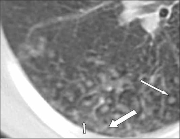
Figure 2.

A twenty-one-year-old man with silicosis. MDCT scan demonstrates ground glass opacity (short arrow), centrilobular nodules (long arrow) and interlobular septal thickening (thick arrow) in the superior segment of the lower lobe of the right lung
