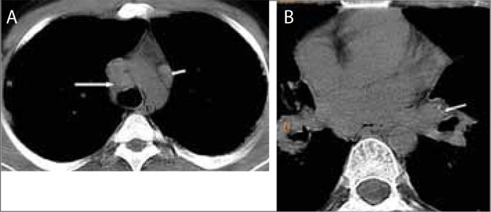
Figure 4.

A twenty-three-year-old man with silicosis. MDCT scan shows lymph nodes larger than 1 cm in short-axis diameter in the paratracheal (long arrow) and prevascular regions (short arrow) (a). MDCT scan shows a lymph node with punctate calcifications in the left hilar region (arrow) (b)
