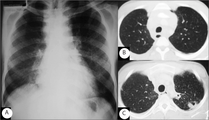
Figure 1.
Chest radiograph shows a reticular pattern in the middle and lower regions of both lungs (A). Axial CT images (lung window) obtained at the level of the upper mediastinum show multiple micronodular opacity in the upper lobe (B) and ill-defined cavities in the left upper lobe (C) on the first and second admission, respectively.

