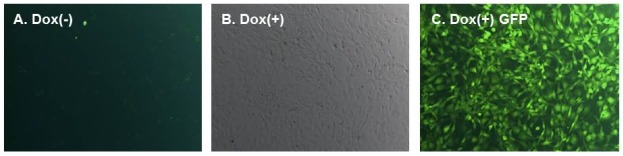
Fig. 1. Microscopic assays for the EGFP expression in hSCP1 inducible cells. Images of hSCP1(Wt and D96N)-induced NIH/3T3 cells in the absence (A) or presence (B) (Phase contrast image) and (C) of 2 μg/ml doxycycline for 72 h were taken using a Leika M205FA stereomicroscope (Leika Microsystems, Wetzler, Germany) equipped with the LAS software for fluorescence imaging.