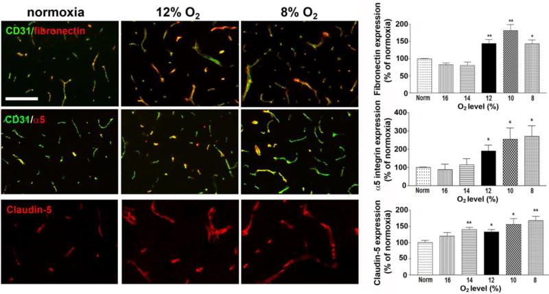
Figure 3.
The influence of hypoxic dose on the expression of angiogenic and tight junction proteins on cerebral vessels. Dual-IF was performed on frozen sections of the frontal lobe from mice exposed to different hypoxic levels (8-16% O2) or normoxia for 7 days using antibodies specific for CD31 or fibronectin, CD31 or α5 integrin, or claudin-5. Scale bar = 100μm. Quantification of the changes in protein expression level are displayed in graphs. All experiments were performed with four different animals per condition, and the results expressed as the mean ± SEM of the % change compared to normoxic conditions. Note that increases in vascular fibronectin and α5 integrin expression were not observed until hypoxic levels reached 12% O2 or lower. In contrast, claudin-5 expression levels were significantly increased at the milder hypoxic level of 14% O2. * P < 0.05, ** P < 0.01.

