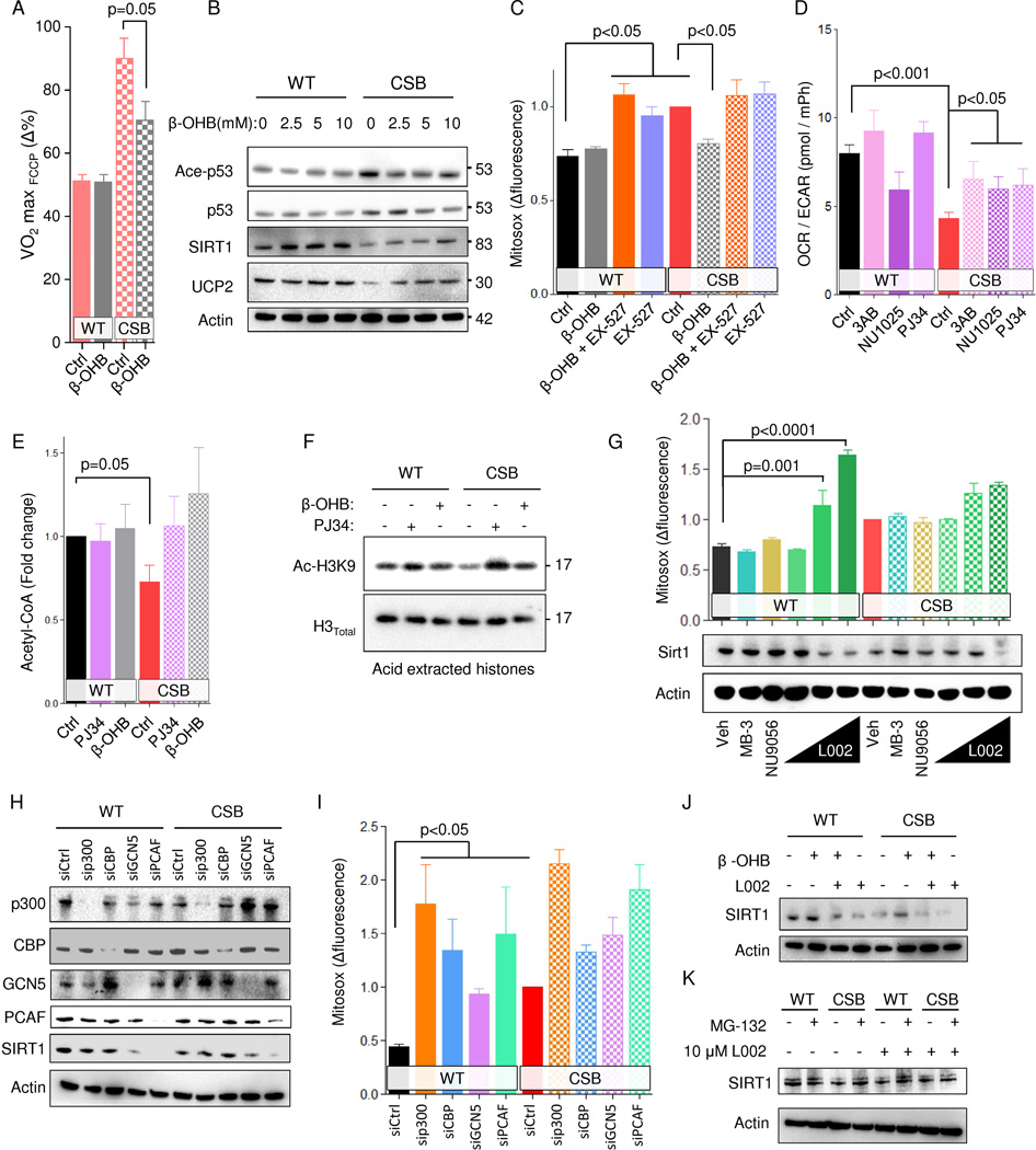
Fig. 4. β-hydroxybutyrate and PARP inhibition rescues the CS phenotype through SIRT1 activation.
(A) FCCP uncoupled respiration normalized to basal respiration 48 hours after 10 mM β-OHB treatment (n=3 separate seahorse experiments, mean ± SEM). (B) Representative immublot of treatment of WT and CSB deficient cells with increasing concentrations of β-OHB. (C) Flow cytometry of WT and CSB deficient cells treated with β-OHB and or EX-527 for 48 hours and stained with mitosox (n=6, mean ± SEM). (D) Oxygen consumption rate (OCR) relative to extracellular acidification rate (ECAR) (n=3–12 separate seahorse experiments, mean ± SEM). (E) Acetyl-CoA levels after treatment with the PARP inhibitor PJ34 or β-OHB for 24 hours (n=6, mean ± SEM). (F) Representative immunoblot of acid extracted histones after treatment with the PARP inhibitor PJ34 or β-OHB for 24 hours. (G) Representative immunoblot from WT and CSB deficient cells after 24 hours treatment with MB-3, NU9056 or increasing concentration of L002 and flow cytometry of WT and CSB deficient cells treated with MB-3, NU9056 or increasing concentration of L002 for 24 hours and stained with mitosox (n=3–12, mean ± SEM). (H) Representative immunoblot of protein levels after knockdown of various proteins. (I) Flow cytometry of WT and CSB deficient cells subjected to siRNA knockdown of various proteins and stained with mitosox (n=3, mean ± SEM). (J) Representative immunoblot of protein levels after treatment with L002 and/or β-OHB. (K) Representative immunoblot of protein levels after treatment with L002 and/or MG-132.

