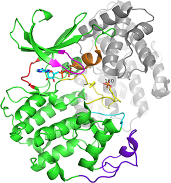
Figure 1.

The structure of the CDK2 - cyclin complex (3QHR [[60]]). This structure is phosphorylated at Thr 160, and bound to cyclin A2 (grey) and ADP (sticks). The PSTAIRE region is shown in orange, the T-loop in yellow, the glycine rich loop in magenta, the CGMC insert in purple, the αEF-αF loop in cyan and the hinge region in red.
