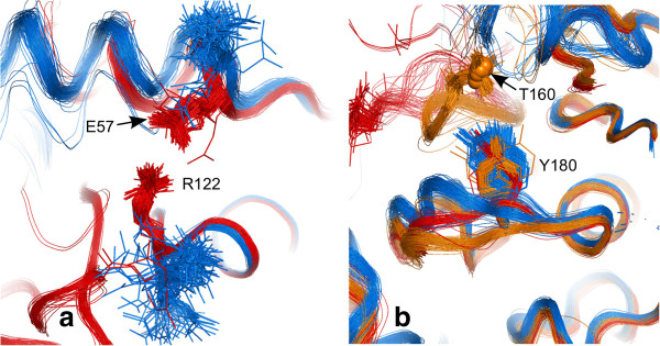
Figure 10.

PCA discriminatory side-chain conformations. (a) salt bridge formation between Arg 122 and Glu 57 in cyclin bound structures (red). (b) Tyr 180 in monomeric (blue), protein-bound but unphosphorylated (red), protein-bound and phosphorylated at Thr 160 (orange, with phosphorus shown as small spheres). The rest of the T-loop is cut away for clarity.
