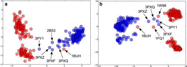
Figure 9.

PCA scores plot for CDK2. Monomeric chains are coloured blue and protein bound chains red. (a) backbone conformation using curvature and torsion. The variance explained by PC1 is 45% and 51% by PC1 and PC2 combined. (b) side-chain conformation using the position of a terminal atom in Cartesian coordinates after transformation of the residue to a reference origin (see Methods). PC1 explains 24% and PC1 plus PC2 explains 35% of the variance. Labels show the PDB codes of outliers.
