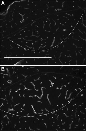
Figure 5.

Testosterone stimulated the growth of the vascular bed in the HVC of female European robins. Photomicrographs of the HVC were prepared from control (A) and testosterone-treated (B) female robins and stained with an anti-laminin antibody. The ventral border of HVC was detected by a DAPI (4′,6-diamidino-2-phenylindole-dihydrochlorid) counterstain (data not shown) and is indicated by a dashed line; scale represents 500 μm.
