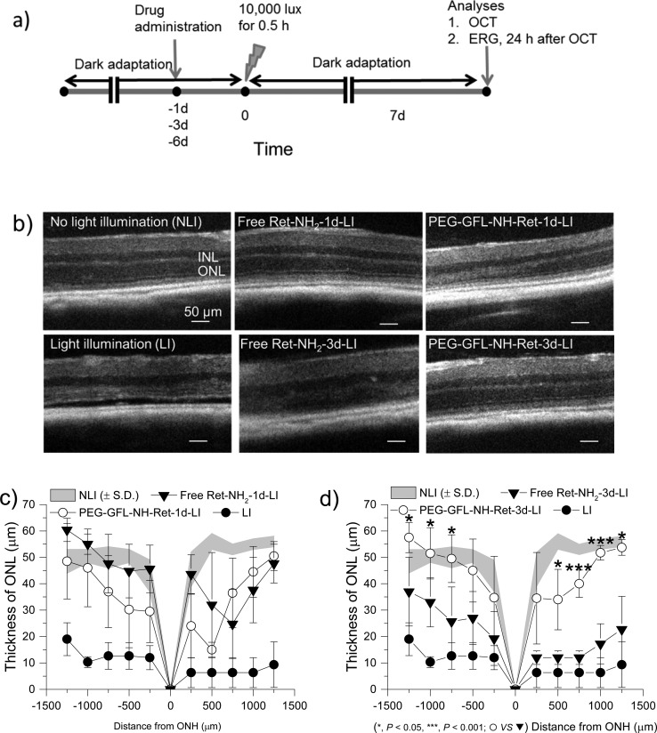
Figure 5.
Protecting effects of PEG-GFL-NH-Ret against light-induced acute retinal degeneration in 4-week-old Abca4–/–Rdh8–/– mice. (a) Schematic representation of the experimental design for assessing the effectiveness of the conjugate. After 4-week-old female Abca4–/–Rdh8–/– mice were kept in the dark for 48 h, they were given either free Ret-NH2 or conjugate PEG-GFL-NH-Ret by gastric gavage at an equivalent dose of 0.5 mg Ret-NH2 per mouse. Mouse eyes were illuminated with 10000 lux light for 30 min either 1 day (1 d), 3 days (3 d), or 6 days (6 d) after the gavage. Mice then were kept in the dark for 7 days, after which final retinal evaluations were performed. (b) Representative OCT images of Abca4–/–Rdh8–/– mouse retinas in different treatment groups (NLI = no light illumination; LI = light illuminated). Scale bar indicates 50 μm in the OCT image. (c, d) Seven days after light exposure, the ONL thickness was measured from in vivo OCT images obtained along the vertical meridian from the superior to inferior retina of mice gavaged either 1 day (c) or 3 days (d) before bright light exposure. Statistical analysis was performed to compare the treatment groups using one-way ANOVA. Error bars indicate SD of the means (n = 4–5).

